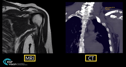
TEL AVIV, Israel & SAN JOSE, Calif.– RSIP Vision, an experienced leader in driving innovation for medical imaging through advanced AI and computer vision solutions, today announces a new tool for Total Shoulder Arthroplasty (TSA) planning. This tool performs segmentation of the shoulder bones from shoulder MRI scan, which is usually performed in shoulder healthcare. The segmentation output undergoes super-resolution enhancement to overcome inherent MRI resolution limitations. The end-result is a high-quality, 3D model of the shoulder bones, which allows exceptional planning for TSA, without the need for a CT scan for planning. This new vendor-neutral technology is available to third-party MRI manufacturers and viewer solutions, allowing an accurate and radiation-free method for TSA planning.
“Shoulder MRI scans are common in shoulder pain management healthcare, usually for soft tissue analysis,” said Ron Soferman, CEO at RSIP Vision. “Deep learning (DL) algorithms can be developed for accurate segmentation of the shoulder bones. Neural networks are trained to process the resulting segmentation into a CT-grade segmentation, improving the original MRI resolution. Further down the line this tool can be altered to segment soft tissue, as well as other anatomies. This tool improves shoulder healthcare as it removes the need for a CT scan and its accompanying radiation and cost.”
Shoulder injuries often require a diagnostic MRI scan, mainly to rule out soft-tissue damage. When approaching TSA, current practice requires a CT scan for procedural planning as CT resolution is superior to that of MRI. However, an additional CT scan involves exposing the patient to harmful radiation, as well as additional healthcare expenses.
RSIP Vision’s new tool utilizes the shoulder MRI scan, without compromising on resolution quality. It automatically segments the humerus and scapula from the scan. The segmentation output goes through another neural network, trained to upgrade segmentation resolution, thus producing a super-resolution model despite the original scan limitations. This output is as-good as CT-based models, without the need for an additional scan, and can be used for procedural planning.
“As a physician, you want to reduce radiation exposure to your patient,” said Dr. Shai Factor, orthopedic surgeon at Tel-Aviv Medical Center. “This new tool by RSIP Vision will utilize existing shoulder MRI scans, which we use routinely to demonstrate associated soft tissue pathologies, and will offer a radiation-free alternative to patients prior to shoulder arthroplasty, without compromising the 3D model’s quality. ”

