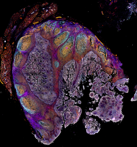
WALTHAM, Mass.– Thermo Fisher Scientific Inc., the world leader in serving science, today introduced the Invitrogen™ EVOS™ S1000 Spatial Imaging System. This advanced system addresses the limitations of current fluorescent microscopy technologies by enabling researchers to generate a multiplexed high-quality image for multiple samples within several hours, thereby lowering the barrier to entry into spatial tissue proteomics.
The EVOS S1000, from Thermo Fisher Scientific’s innovative line of cell imaging microscopes and systems, leverages advanced and patented spectral technology, allowing researchers to capture images of up to 9 different targets simultaneously, which helps reduce the need for multiple rounds of imaging and preserves tissue integrity.
“Understanding tissue structure and function is crucial for developing new treatments for solid tumors and neurodegenerative diseases,” said Trisha Dowling, vice president and general manager for flow and imaging technologies at Thermo Fisher Scientific. “The EVOS S1000 delivers a detailed snapshot of tissue microenvironments and architecture in their native state, helping researchers accelerate their experiments, achieve more with their tissue samples and drive advancements in critical research areas.”
The system’s compatibility with a wide range of reagents and antibodies enables seamless integration into existing laboratory setups, helping meet the growing demand for multiplex imaging.
“Our lab handles everything from project design and sample preparation to imaging, and it’s critical that we deliver high-quality results to our customers, while also working to support our own research projects,” said Carolina Oses Sepúlveda, researcher and lab manager for spatial proteomics at SciLifeLab Stockholm. “With the new EVOS S1000, we can select and utilize any antibody or reagent – allowing us more flexibility to choose the tools that best fit our research needs – or even work without antibodies, all of which reduces sample processing time.”


