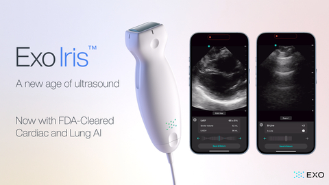
SANTA CLARA, Calif.– Exo® (pronounced “echo”), a medical imaging software and devices company, today announced its FDA-cleared cardiac and lung artificial intelligence (AI) applications are now available on Exo Iris™, Exo’s high-performance handheld ultrasound device.
With affordable, AI-powered medical imaging in a pocket-sized device, caregivers can get to answers immediately to accelerate diagnosis and create new care pathways for heart failure patients at the point of care. As an industry leader in AI, Exo now has FDA 510k clearances on five different applications—cardiac, lung, bladder, hip and thyroid—with plans to double the number of clearances by 2025.
The number of Americans with heart failure today is over 6.7 million and growing. This figure represents both a population health and financial crisis with annual healthcare spending ballooning to $240 billion by 2030*. While echocardiography is the cornerstone of heart failure diagnosis, it is costly and requires highly trained sonographers to use it. As a result, people go without medical imaging when and where they need it most.
AI-powered handheld ultrasound can lower the threshold for answers at the point of care, enabling access to simple and affordable medical imaging. With the launch of Exo’s latest AI capabilities, health systems and caregivers—particularly those in rural and under-resourced settings—can leverage Exo Iris to simplify the process of obtaining and interpreting medical images specific to the heart and lungs. As a result, caregivers can accelerate their patients’ diagnosis and treatment no matter where they are located.
“It’s time for a reimagined approach to addressing heart failure at scale. That’s why Exo is putting AI-empowered medical imaging in the hands of every caregiver, no matter their specialty,” said Sandeep Akkaraju, Co-founder and CEO of Exo. “Exo’s AI is simple to use, reproducible and objective, and will contribute to more accessible and reliable healthcare for all.”
Exo trains its AI applications on over 100,000 images from point-of-care ultrasound (POCUS) exams performed by users in real-world settings, including emergency rooms and critical care units. As part of the FDA clearance process, Exo’s AI was rigorously validated across various patient populations, health conditions and diagnostic scan types. Therefore, Exo’s AI recognizes internal landmarks even on less-than-perfect scans, enabling caregivers to gain the real-time data needed to make informed decisions without requiring lab-quality ultrasounds.
With AI designed around the caregiver, questions like “Is my patient’s shortness of breath related to a heart condition or a lung condition?” can now be answered in a matter of seconds. With a quick scan, Exo’s lung AI reliably identifies the presence of B-lines, enabling users to quickly assess if the patient has pulmonary edema, or fluid in the lungs. Also, both expert and early POCUS users can leverage Exo’s cardiac AI to quickly measure left ventricle ejection fraction (LVEF) and stroke volume in a few heartbeats in both parasternal long axis (PLAX) and apical four-chamber (A4C) views. All these valuable data can be acquired and interpreted in real-time so that clinicians can improve patient care.
“Exo’s cardiac and lung AI applications are a game-changer,” said Ted Koutouzis, Clinical Instructor in Emergency Medicine at Northwestern Medicine. “Now I have a fast and reliable clinical tool to help easily distinguish between COPD and CHF patients for precise and timely care. What’s even more exciting is how these applications open ultrasound technology to a whole new set of POCUS users, especially those with limited experience. When clinicians can perform medical imaging efficiently, it’s a win-win for both the patients we serve and the healthcare systems we work within.”
In addition to cardiac and lung AI, Exo Iris now features Pulsed-Wave Doppler capabilities to provide physicians with even more opportunities to look at blood velocity to support diagnosis and deeper findings in cardiac, abdominal and vascular applications.


