ATLANTA– Vektor Medical, a leader in non-invasive, AI-powered arrhythmia analysis technology, will present findings today at HRX 2024. The two studies showcase how Vektor’s advanced technologies uniquely leverage AI and deep learning to assess wall thickness and scar tissue in ventricular tachycardia (VT) patients, providing crucial insights for electrophysiologists performing ablation procedures. Both studies will be featured during the Top 5 AbstracX session at 2:45 PM, highlighting the highest-scoring abstracts submitted to HRX 2024.
In addition to these studies, on the HRX Main Stage, Dr. Anish Amin, System Medical Chief for Cardiac Electrophysiology and Medical Director of the Atrial Fibrillation Clinic at Riverside Methodist Hospital, will discuss how vMap addresses post-PVI and PFA reconnection challenges and is shaping the future of arrhythmia care. His presentation, titled “Beyond PVI: Redefining the Future of Arrhythmia Management,” will emphasize personalized AF interventions, broader access to advanced therapies, and innovative strategies for managing complex arrhythmias.
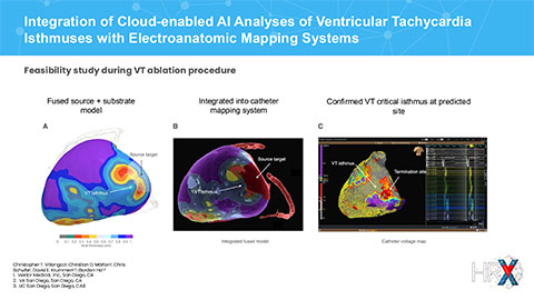
The first study, Integration of Cloud-enabled AI Analyses of Ventricular Tachycardia Isthmuses with Electroanatomic Mapping Systems, demonstrates a seamless integration of vMap’s ECG analysis with AI-driven CT substrate analysis. This innovative approach fuses ECG and CT data to deliver a multimodal analysis of VT source locations and arrhythmia substrates. The study analyzed the ECGs of 31 ventricular tachycardia patients using computational mapping, which was then combined with corresponding CT scans on Vektor’s cloud-based platform. An ensemble of deep-learning models generated detailed masks of cardiac chambers, meshes, and LV myocardial thicknesses, which were then co-registered with ECG-localized VT exit sites. These fused models were imported into electroanatomic mapping systems for precise catheter guidance during VT ablation procedures.
Study Conclusion: Vektor’s cloud-enabled AI analysis of CT and 12-lead ECG provides a rapid, end-to-end solution for accurately localizing VT isthmuses within scar tissue, correlating with invasive activation mapping and VT termination sites. This method facilitated precise and efficient VT ablation.
A second study, Validation of a Deep Learning Ventricular Tachycardia Substrate Model, evaluated the efficacy of AI algorithms in enhancing the analysis of myocardial wall thickness – an emerging technique in identifying arrhythmogenic tissue underlying VT. Vektor’s AI-based process utilized raw CT scans to create a three-dimensional wall-thinning heart model, accurately segmenting cardiac structures in 31 VT patients with various heart morphologies and imaging artifacts.
Key Data Points & Conclusion: The AI model achieved high accuracy in segmenting cardiac structures in VT patients, with wall thinning measurements aligning closely with invasive substrate mapping. The model achieved mean Dice-scores of 0.96 ± 0.02 (LV), 0.86 ± 0.03 (MYO), 0.94 ± 0.01 (LA), 0.87 ±0.04 (RA), 0.91 ± 0.03 (RV), 0.92 ± 0.02 (Aorta), 0.91 ± 0.02 (Pulmonary Artery), 0.79 ± 0.07 (Atrial Appendage Left), 0.70 ± 0.11 (Esophagus), 0.69 ± 0.19 (Inferior Vena Cava), 0.76 ± 0.04 (Pulmonary Vein), and 0.85 ±0.05 (Superior Vena Cava).
“AI technology has long promised to revolutionize healthcare, and today, we’re seeing that promise become reality,” said Rob Krummen, CEO of Vektor Medical. “The results of these studies underscore how Vektor Medical continues to push the boundaries of what is possible with AI in arrhythmia care. I’m incredibly proud of our team for earning two of the top five abstracts at HRX 2024. Our company is committed to streamlining procedures, enhancing clinical efficiency, and, above all, improving patient outcomes. We are excited to share these findings with the passionate professionals dedicated to transforming healthcare at HRX 2024.”


